Congnos Therapeutics
Unlocking the Future of Therapeutic Health
Discover a transformative therapeutic platform that reshapes the current standard of neurological and oncological care – a step towards a healthier, happier lifestyle.


Cognos Mission
Leading the convergence of science, data and technology utilizing AI and Blockchain
At Cognos Therapeutics, our mission is to develop and commercialize medical products that combine diagnostic, therapeutic, and sensing technologies with state-of-the-art drug delivery to advance healthcare through improved patient outcomes.

A Novel and Ground-Breaking Advancement
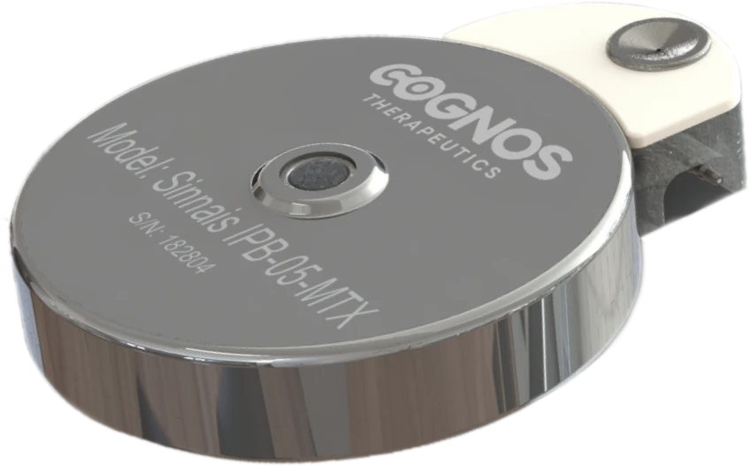
SINNAIS™, Implantable Smart Pump
Our innovative SINNAIS™ implantable pump represents a platform designed for the future, utilizing power of AI, blockchain and cloud connectivity and incorporating microprocessor, microelectronics, communication capabilities, and embedded programmable parameters in its architecture. Focusing on targeted, controlled delivery of therapeutics to the brain a departure from the current standard of intraventricular drug delivery.
Target indications
Serving Unmet Needs
$15.8B
Combined Treatment Market Size
Treating central nervous system (CNS) diseases, including multiple neurological and other diseases such as cancer, is difficult because the blood-brain barrier, which helps protect the brain from viral and bacterial infections, also prevents the penetration of most drugs at effective therapeutic levels.
180K
Total Addressable Market (TAM)
The total addressable market size for SINNAIS™ in the United States is at least 180,000 patient cases per year, comprised of estimated annual diagnoses (i) of 110,000 for Leptomeningeal carcinomatosis (LC) cancer, (ii) 60,000 for pancreatic cancer, and (iii) 12,000 for glioblastoma. Within the LC diagnoses, breast cancer, melanoma, and lung cancer make up more than 50% of the potential target population.
We estimate the combined treatment market for these three diagnoses in the United States at approximately $15.8 billion per year, comprised of approximately $6 billion for LC, approximately $8 billion for pancreatic cancer and approximately $1.8 billion for glioblastoma.
$15.8B
Combined Treatment Market Size
180K
Total Addressable Market (TAM)